
Introduction: Beetle Dissection and Mounting


In order to make identifications of rove beetles and especially for Aleocharinae (1-9mm long) (Coleoptera, Staphylinidae, Aleocharinae), genitalia have to be dissected and mounted. These beetles are extremely abundant and are found in soil litter and mushrooms in diverse environment. There are currently over 400 species of Aleocharinae described in Canada. The collected specimens are preserved in alcohol and need to be dissected to examine the genitalia for identification. The following steps will show you how to perform a beetle dissection and permanent mount to examine the genitalia.
Step 1: Materials List

Material list
- 2 fine soft tweezers
- 2 needle holders
- a double well glass plate
- Pinning plate
- pinning block
- dissecting stereoscope and light
- Pins ( #3 preferable),
- mounting card (4x11mm),
- celluloid microslides,
- Canada balsam,
- water,
- absolute alcohol,
- water soluble glue,
- beetle specimens from various sources
Prepare the insect pins by putting the card onto the pin using the pinning block to adjust the height of the card; then the microslide underneath. Put a drop of Canada balsam on the transparent micoslide. Put the pin aside on the pinning plate ( the prepared pins are on the pinning plate).
Step 2: The Specimen


Take a specimen ( here a 2 mm Gyrophaena ). Put a drop of water in a well on the glass plate and deposit the beetle specimen
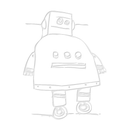
Step 3: Basic Schematic of a Beetle

Here is a simple schematic of a beetle to show you the various part of the beetle. the dissection is performed by inserting a needle between the 7th and 8th segment and separate the two
Step 4: Separate the Terminal Segment


Under the dissecting scope with a 30 to 45X magnification, hold the beetle with a tweezer while with the other hand insert the needle between the 7th and 8th segment and separate the two .
- Separate the 8th tergite from the 9th terminal segment using two needles;
- separate the tergite from the sternite (the other side of the segment).
- The genitalia are encapsulated between these segments
- Remove the beetle from the well unto an absorbing paper
Step 5: Separate the Genital Parts

If you have a female aleochrinae you will find a single tube (spermateca) while the male will have a tube enclosed between two clasps. Separate the clasp (paramers) from the tube (aedeagus) ( the needle here is holding the aedeagus and paramers)
Step 6: Placing the Insect Parts on the Canada Balsam


All the 3 beetle parts are now transferred to a separate well containing absolute alcohol using the tweezers, to dehydrate the parts before transferring them on the Canada balsam on the mounted microslide pin.
Once in the Canada balsam, make sure all parts are covered by the Canada balsam, remove all air bubbles and position them consistently (e.g. tergite, sternite, terminal segment on the left, genitalia on the right); the needle is showing the aedeagus in the enlarged picture
Step 7: Mount the Insect on a Card


Place a minute drop of water soluble glue on the card. Place the dry beetle on the glue and position the antennae, legs, and body so that all body parts are well displayed for identification
Let dry the Canada balsam for 2-3 weeks and then insert labels containing the data for the specimen regarding the locality and geographical position ( lat, long, elevation), date of collection, habitat data, collection method and collector name.
Your specimen can now be handled for identification.
Step 8: Pinned Insect Box - Ready for Identification

Repeat several hundred times!